Laurie says:
..
Making my Pandemic Shadow images has definitely made me much more aware of small nuances. These photographs would have attracted me anyway, but at this time I’m far more fascinated by smallness. And these images let us into worlds that we would never see otherwise.
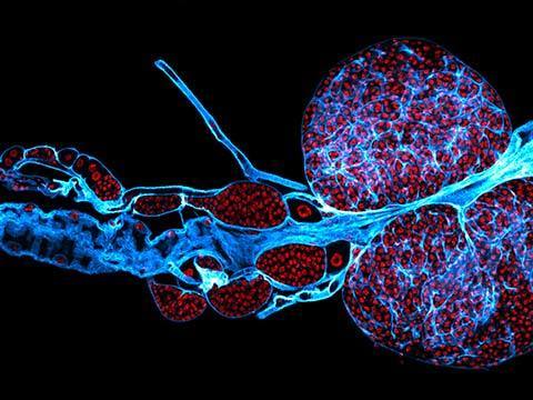
The image above won first place for 2020 in the Nikon Small World contest this year. It’s a dorsal view of bones and scales (blue) and lymphatic vessels (orange) in a juvenile zebrafish. It’s by Daniel Castranova, Dr. Brant M. Weinstein, and Bakary Samasa. The magnification is 4x. The colors and textures are superb.
..

..
This took 4th place. It was of Multi-nucleate spores and hyphae of a soil fungus (arbuscular mycorrhizal fungus). It is the work of Dr. Vasileios Kokkoris, Dr. Franck Stefani, and Dr. Nicolas Corradi. The magnification is 63x. I love the complexity and the contrasts of the image.
..
This took 6th place. It’s a Hebe plant anther with pollen. It is the work of Dr. Robert Markus and Zsuzsa Markus. The magnification is 10x. I think it has an intense textural quality that’s almost fabric-like.
..
This photograph was an Honorable Mention. It’s the Lymph gland (blood organ) of a fruit fly larva. It is the work of Dr. Saikat Ghosh and Dr. Lolitika Mandal. The magnification is 40x. It’s just gorgeous.
I’m grateful for the opportunity to see into what would otherwise be unknown worlds. Explore the worlds.
Follow Debbie on Twitter.
Follow my new Pandemic Shadow photos on Instagram
![]()


